Types of investigations

Echocardiography
Echocardiography is a type of ultrasound used to assess heart chambers’ size and function and it looks for structural abnormalities in the way how valves work. Depending on what information is needed, you may need one of several types of echocardiograms (transoesophageal echo, bubble study, contrast echo, or stress echo using either exercise or pharmacological stress to assess the supply of oxygen to different parts of the heart).
If you have a significant problem with the function of one of the valves Dr Wamil may suggest transesophageal echo to acquire more-detailed images which are necessary to guide surgical treatment. It is difficult to get a clear picture of your heart valves with a standard echocardiogram. TOE will allow Dr Wamil to use 3D reconstructions of images to get a very clear assessment of the mechanism of disease and suggest the most appropriate way of treatment.
Dr Wamil completed sub-speciality training in cardiac imaging in the Oxford University Hospitals and has accreditations in echocardiography and transoesophageal echocardiography issued by the British Society of Echocardiography and the European Association of Cardiovascular Imaging.
Dr Wamil is the co-author of a chapter about TOE in a very popular book for cardiologists.
She also published popular educational articles (1) (2) describing the technique in more detail.

Cardiac CT coronary angiography
Cardiac CT coronary angiography uses low radiation to assess the coronary arteries that supply blood and oxygen to the heart muscle in a non-invasive way. That technique is also used to plan structural heart disease procedures such as TAVI (a replacement of the aortic valve), MitraClip (a treatment for the mitral regurgitation) and can assess the grafts after the bypass operation (CABG). Dr Wamil’s training in CTCA has been certified by the Society of Cardiac Computed Tomography.
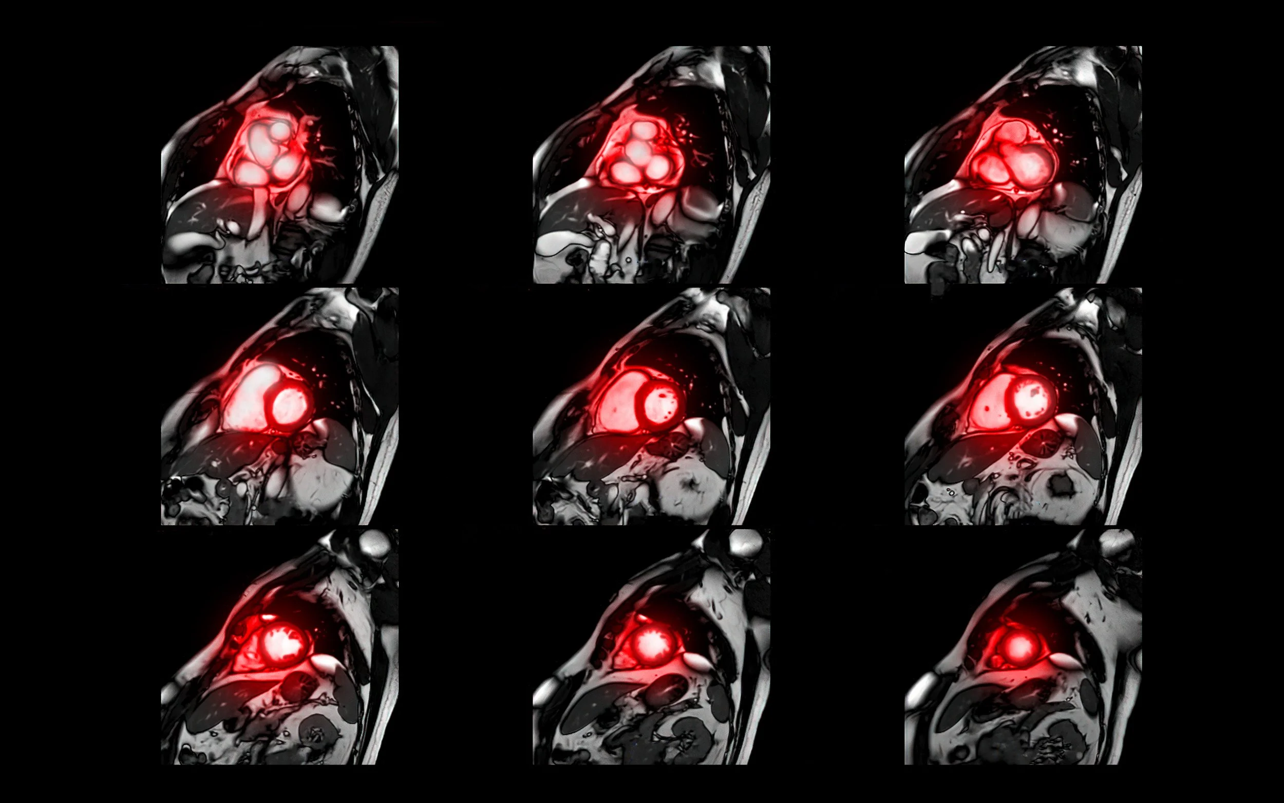
Cardiac magnetic resonance (CMR)
Cardiac magnetic resonance (CMR) allows cardiologists to view exceptionally detailed images of the heart structure and blood vessels. A CMR scan helps to see how the heart is functioning and will assist Dr Wamil in gaining a better understanding of the heart disease that caused your symptoms. Using various scanning protocols Dr Wamil can evaluate cardiac function, heart muscle scarring, problems with poor blood supply, and assess certain types of valve problems. Dr Wamil has expertise in using novel CMR techniques such as T1 and T2 mapping in assessing causes of heart failure and rare cardiomyopathies. She has level 3 accreditations in CMR from the European Association of Cardiovascular Imaging and from Society of Cardiac Magnetic Resonance.
How multimodality imaging is helping patients

Imaging plays an essential role in the diagnosis and management of heart diseases. A key concept in multimodality imaging is personalised patient-centered approach to select the best available imaging test when evaluating patients with known or suspected heart disease. While many clinical questions can be answered by a single test, the effective use of multimodality cardiac imaging requires collecting information from different imaging scans. Such an approach decreases the patient’s risks of using various techniques and strengthens the evidence required to make an accurate diagnosis and propose the most appropriate course of treatment.
Dr Wamil is one of a very few cardiology consultants in the UK who has expertise in three cardiac imaging modalities (echo, cardiac CT and CMR) and therefore can deliver truly personalised management of heart diseases carefully tailored to all patients needs.
Here, Dr Wamil discusses the use of multimodality imaging in the management of coronary artery disease.
Here, Dr Wamil discusses the use of multimodality imaging in the management of coronary artery disease.
Role of Non-invasive Cardiac Imaging in the Management of Coronary Artery Disease